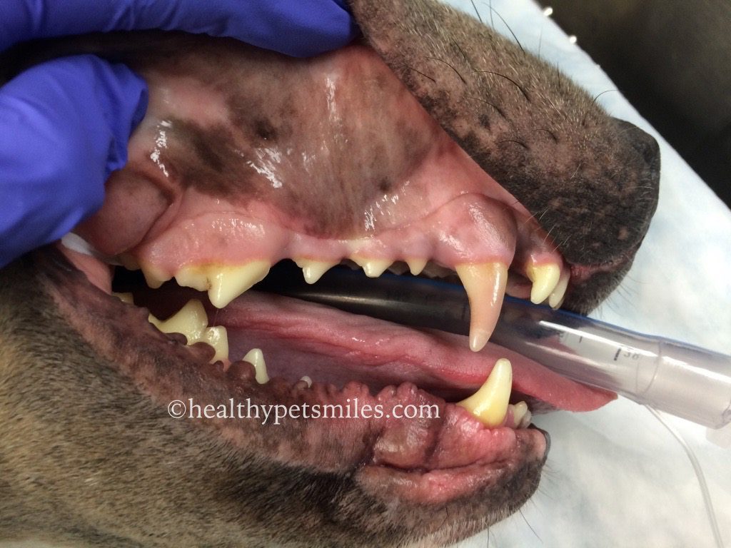
Discolored Teeth
In a veterinary practice, it is not uncommon to see discolored teeth in our patients. There are many causes of discoloration, including trauma, infection, metabolic disease, or drug-induced. When evaluating discolored teeth, many things should be taken into consideration including age, history, overall health, and how long the tooth/teeth have been discolored. All of these are important factors in determining the cause of the discoloration and the potential treatment options. Discolored teeth are evaluated both visually and radiographically.
Diagnosis:
Diagnosis of discolored teeth starts includes visual examination and radiographs as detailed below.
Visual examination:
Visual inspection of a discolored tooth often starts with a procedure called transillumination. A bright light source is directed at the tooth surface/crown. If the pulp of the tooth is alive and healthy, the tooth will glow a pinkish hue. However, if the pulp is non-vital, the crown will glow a dull yellow or grey color and the light will not shine through.
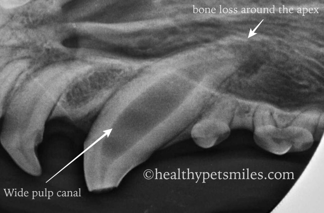
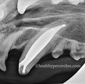
X-rays:
Radiographs are a valuable means of fully evaluating discolored teeth. On x-rays, we are looking at several key elements to see if the tooth shows signs of internal disease (endodontic disease). The criteria evaluated includes the width of the root canal, the health of the bone around the apex of the tooth, and the width and presence of the periodontal ligament. If the root canal is a different width than those of the other teeth around it, or if there is evidence of bone loss around the apex of the tooth, these are indicators of endodontic disease and treatment should be initiated immediately via root canal therapy or extraction. It is important to note that x-rays will not show signs of early disease, so it is possible that your primary veterinarian or veterinary dentist might recommend treatment even if the x-ray findings are unremarkable.
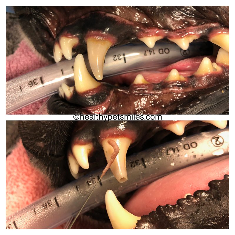
Treatment:
In previous studies, it has been found that the vast majority of discolored teeth are non-vital, so regardless of visual and radiographic findings, treatment is often recommended for these teeth. The two treatment options available are root canal therapy or extraction, both of which have good long-term success. It is important to discuss these options with your veterinary care provider.
Consequences of failure to treat non-vital teeth:
Non-vital teeth invariably have a necrotic pulp with serves as a potential nidus for local infection (periapical or osteomyelitis) or systemic infection (ex: heart, lung, kidney). Additionally, periapical pathology associated with non-vital teeth is painful! Have you ever experienced a toothache? These personal experiences can directly affect attitudes with regard to dental care for dogs and cats. Because our dogs and cats rarely alert us to a painful problem in their mouth, we must remain vigilant to ensure that they have pain-free mouths.